By sarms4muscle.com | 08 January 2024 | 0 Comments
Immune Cells in the Obesogenic Environment
Adipose tissue (AT), including visceral adipose tissue (VAT) and subcutaneous adipose tissue (SAT), is a source of inflammatory cytokine production.
AT influences both inflammatory and adaptive immune functions. Obesity leads to the accumulation of pro-inflammatory immune cells (including macrophages and lymphocytes) in visceral adipose tissue, which have high levels of nutrient metabolism that drive low-grade inflammation and promote metabolic disease.
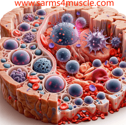
Immune cells in adipose tissue

Immune cells in adipose tissue
B cells and their antibodies
Primarily pathogenic IgG antibody production. B-lymphocyte infiltration was observed in the VAT of diet-induced obese mice, producing pathogenic IgG antibodies.
This adipocyte-specific IgG antibody secretion results from the ongoing adipocyte cell death process in the obese AT, which induces the release of chronically stimulated "self" antigens from B cells. This process occurs without exogenous stimulation.
Most of the IgG produced is concentrated in the coronal region of the VAT and consists of large mononuclear and multinucleated macrophages and dying adipocytes, which may be involved in the clearance of dying adipocytes.
In humans, this pathogenic IgG secreted by B cells expresses GOSR1, BTK, GFAP, and HSP60, the first three of which are closely related to their insulin resistance, mostly intracellular and widely expressed.
APC.
Dying adipocytes elicit humoral responses against autoantigens expressed by AT cells. Adipocytes express typical markers of antigen-presenting cells (APCs), such as CD1d and MHCII. characterized according to the antigenic specificity of the autoantibodies, and then categorized according to the cellular location of the antigen (nearly half of them are cytoplasmic) and the biological processes in which the antigen is involved (mainly metabolic processes).
DC: is the major APC accumulated in obese mice and human AT, inducing Th17 cell differentiation. Compared to normal DCs, DCs in obese tissues express low levels of CD40, CD80, CD86 and MHC. Lack of CD40 was associated with weight gain and degree of insulin resistance. In addition, conventional ATDC express CD11c+ CD64- and are dependent on CCR7 for proliferation and accumulation in a high-fat dietary environment.
Macrophages: CD40 is an influential factor co-expressed by DCs and macrophages, but levels are diametrically opposed in both APCs.
Mouse models show high expression of CD40 by macrophages in obesity AT. knockout of CD40 in mice resulted in reduced expression of MHCII and CD86 on macrophage membranes, but was not associated with improved insulin sensitivity. This also explains why CD40 has an opposite role in AT inflammation.
T cells
T cells in AT express more IFN-γ+ and GZMB+ and are able to produce more inflammatory mediators upon activation of TCR signaling.
CD4+ T cells are able to modulate obesity-related metabolic abnormalities by a mechanism that does not involve Treg cells or IL-10 but rather Th2 cells to address metabolic deficits such as visceral adiposity and insulin sensitivity caused by adipocyte hypertrophy.
The ratio of T cell subsets in AT is also different from that in normal tissues. For example, the proportion of naïve T cells is reduced in SAT, while effector T cells and memory T cell subsets are increased in VAT.
Th1/Treg: The overall Treg ratio is low in obese AT.The Th1/Treg ratio is affected by the STAT3 signaling pathway.
The Treg ratio is lower in VAT than in SAT, while Th1 cells are more abundant than in SAT. A high-fat diet increases the proportion of Th1 in VAT and thus the Th1/Treg ratio, and BMI is negatively correlated with Treg values in VAT.
CD8+ Memory T Cells: A special group of CD8+ memory T cells also exists in SAT with high expression of metallothionein genes (MT1E, MT1F, MT1G, MT1X, and MT2A). This subpopulation is able to increase the expression of IL-2 receptors on T cells while inhibiting T cell differentiation towards cytotoxic lymphocytes, leading instead to an increase in the proportion of naïve T cells and a decrease in the cytotoxic effector T cell phenotype.
Th17 cells promote preadipocyte differentiation and pro-inflammatory cytokine production by producing IL-17, resulting in AT inflammation. In VAT, IL-17 increases the number of Th1 cells and decreases the number of Th2 cells and M2 macrophages mediated through TBK1.
iNKT cells
The role of iNKT, which recognizes lipid antigens expressed on M2 macrophages and adipocytes presented by CD1d, on obese body weight and glucose metabolism is complex.
In obese human and mouse AT it was observed that iNKT was depleted, increased in parallel with pro-inflammatory macrophage infiltration, and recovered after weight loss.
Positive effects: adipose-retained iNKT produced less IFN-γ and more IL-4 and IL-10 in the liver and spleen. blockade of IL-4 and IL-10 secretion attenuated the effects of iNKT activation on amelioration of insulin resistance, but the effects on weight loss remained unchanged.
Positive effects of iNKT on body weight and glucose metabolism encompassed secretion of fibroblast growth factor 21 (FGF-21), which in turn induced browning of white adipose tissue (WAT) through up-regulated coupling protein 1 (UCP1) expression, as well as polarization of macrophages through production of IL-2 and IL-10 by Treg and recruitment and M2 macrophages.
Negative effects: in other mouse experiments, however, it was observed that iNKT was increased in the AT of obese mice, and that the high-fat diet induced the secretion of pro-inflammatory cytokines but not anti-inflammatory cytokines by iNKT.
NK cells.
Both NK cells and NK cell-produced IFN-γ levels were higher in VAT of diet-induced obese mice than in lean mice due to obesity-induced up-regulation of adipocyte-surface NK cell ligands.NK cells and IFN-γ, in turn, promote macrophage differentiation into pro-inflammatory M1 phenotypes and influence insulin resistance.
Macrophages.
Macrophages are recruited to VAT and SAT during obesity, and their numbers correlate with adipocyte size and body weight. Macrophage infiltration is associated with lipolysis of adipocytes and the formation of multinucleated giant cells. Of the top 100 genes associated with weight gain, 30 encode macrophage-associated proteins.
Macrophages in the AT of lean mice consisted predominantly of M2-polarized cells; in contrast, the M1/M2 ratio was increased in diet-induced obese mice.
Neutrophils, eosinophils, mast cells
Their main role is to influence macrophage recruitment and infiltration.
Neutrophils interact with adipocytes through CD11b and ICAM-1.
Eosinophils also play an important role in macrophage recruitment in AT, acting as a major source of IL-4 to activate macrophages and inducing their differentiation toward the M2 phenotype.
Mast cells promote inflammation, apoptosis, and angiogenesis in the AT by secreting IL-6 and IFN-γ, which contribute to obesity and impaired glucose tolerance. Obese humans and mice have higher numbers of mast cells than lean individuals. Mast cell membrane stabilizers improve body weight and insulin sensitivity in obese mice.
Puma New Drug Alisertib 1028486-01-2
A Paradigm Shift in Targeted Therapy for HR+/HER2- Advanced Breast Cancer
Leave a Reply
Your email address will not be published.Required fields are marked. *